| Mar 21, 2024 |
|
|
|
(Nanowerk News) The Interdisciplinary Research Institute of Grenoble (CEA-Irig), CEA-Leti and fellow European and Canadian institutes and researchers have demonstrated the complete vascularization of organoids on a microfluidic chip at speeds and flow rates similar to blood’s, improving functional maturation and enabling their long-term survival.
|
|
Organoids, which are a 3D assembly of self-organizing cells capable of partially mimicking different physiological characteristics of an organ or tissue, are proving to be highly useful for evaluating the therapeutic efficacy of drugs or new molecules. But they must be vascularized to promote the exchange and transport of nutrients and oxygen, otherwise their maturation and growth are impaired. In vivo, this vascularization is ensured by blood flow.
|
|
By vascularizing organoids in vitro and maintaining them in culture for 30 days in a microfluidic chip, researchers observed significant improvement in their growth, maturation and physiological functions, virtually equivalent to those observed after xenotransplantation in mice. This significant technological advance in organoid R&D also enables production scaling.
|
 |
| ISO-conform microfluidic systems made of stable thermoplastics by micromachining and hot embossing for the organoid culture and vascularization. (Image: CEA-Leti)
|
|
The breakthrough was reported in Nature Communications (“A microfluidic platform integrating functional vascularized organoids-on-chip”).
|
|
“The development of vascular networks in microfluidic chips is crucial for the long-term culture of three-dimensional cell aggregates such as spheroids, organoids, tumoroids, or tissue explants,” the paper explains. “Despite rapid advancement in microvascular network systems and organoid technologies, vascularizing organoids-on-chips remains a challenge in tissue engineering. Most existing microfluidic devices poorly reflect the complexity of in vivo flows and require complex technical set-ups.”
|
|
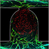
The team’s innovative idea was first to develop a self-organizing vascular network within the chip and then trap an organoid containing its own endothelial cells within it. Both networks are self-connected and they enabled the organoid to be perfused in vitro, mimicking the blood system.
|
 |
| Endothelial network formation perfusing an organoid trapped in the microfluidic chip. (Image: CEA-Leti)
|
|
“This work opens new avenues to understand biological mechanisms in much more relevant models of human origin, as well as for drug discovery and drug development of novel biological therapies,” said Xavier Gidrol, CEA-Irig scientist and project supervisor. “Organoids have now entered the field of personalized medicine, regenerative medicine and pharmacological research.”
|
|
“We have demonstrated a never-reported, improved functional maturation of the vascularized organoid-on-chip by using a reliable microfluidic chip made of thermoplastics, which are well-known in the plastic industries and compatible with production scaling in the near future,” said Fabrice Navarro, a CEA-Leti scientist and co-author of the paper.
|
|
The project included scientists and research engineers from France, Austria and Canada.
|


