| Sep 03, 2024 |
|
(Nanowerk News) As far as water gear goes, floaties are not exactly high tech. But the tiny air-filled bubbles some microorganisms use as flotation devices when they compete for light on the water surface are a different story.
|
|
Known as gas vesicles (GVs), the micrometer-sized bubbles hold great promise for a host of biomedical applications, including imaging, sensing, cellular manipulation and tracking and more. The problem is researchers do not yet know how to make medically useful GV varieties in the lab.
|
|
Rice University bioengineers have now created a road map showing how a group of proteins interact to give rise to the bubbles’ nanometer-thin shell. By detangling some of the complex molecular processes that take place during GV assembly, Rice bioengineer George Lu and his team in the Laboratory for Synthetic Macromolecular Assemblies are now one step closer to unlocking powerful new diagnostics and therapeutics based on these naturally occurring structures.
|
|
“GVs are essentially tiny bubbles of air, so they can be used together with ultrasound to make things inside our bodies visible such as cancer or specific parts of the body,” said Manuel Iburg, a Rice postdoctoral researcher who is the lead author on a study published in The EMBO Journal (“Elucidating the Assembly of Gas Vesicles by Systematic Protein-Protein Interaction Analysis”). “However, GVs cannot be made in a test tube or on an assembly line, and we cannot manufacture them from scratch.”
|
 |
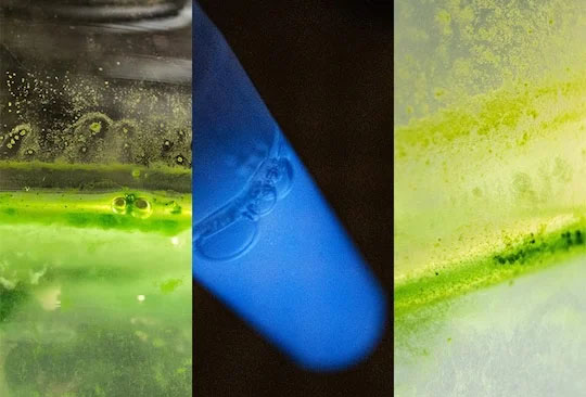
| The tiny air-filled bubbles some photosynthetic microorganisms use as flotation devices could be engineered into powerful biomedical applications. Rice University bioengineers created a road map of the protein-protein interactions that give rise to the formation of these gas vesicles in microorganisms. Part of the process involved using bioluminescence as a tool for gauging protein-protein interactions. (Image: Jeff Fitlow, Rice University)
|
|
The family of GVs includes some of the smallest bubbles ever made, and they can subsist for months. Their stability over longer durations is due largely to the special structure of their protein shell, which is permeable to both individual water and gas molecules but has an inner surface that is highly water-repellent ⎯ hence the GVs’ ability to keep gas in even as they are submerged. And unlike synthetic nanobubbles, which are supplied with gas from without, GVs harness gas from the surrounding liquid.
|
|
The water-dwelling photosynthetic bacteria that use GVs to float closer to sunlight have specific genes encoding for the proteins that make up this special shell. However, despite knowing just how the tiny bubbles look and even why they tend to cluster together, researchers have yet to figure out the protein interactions that enable the structures’ assembly process. Without some insight into the workings of these protein building blocks, plans for deploying lab-engineered GVs in medical applications have to be placed on hold.
|
|
To address the problem, the researchers honed in on a group of 11 proteins they knew were part of the assembly process and figured out a method to track how each of them, in turn, interacts with the others inside the living parent cells.
|
|
“We had to be extremely thorough and constantly check whether our cells were still making GVs,” Iburg said. “One of the things we learned is that some of the GV proteins can be modified without too much trouble.”
|
|
The researchers used this insight to add or subtract certain GV proteins as they were running the tests, which allowed them to figure out that interactions between some of the proteins required help from other proteins in order to unfold properly. They also checked whether these individual interactions changed over the course of the GV assembly process.
|
|
“Through many such permutations and iterations, we created a road map showing how all these different proteins have to interact to produce a GV inside the cell,” Iburg said. “We learned from our experiments that this road map of GV interactions is very dense with many interdependent elements. Some of the GV proteins form subnetworks that seem to perform a smaller function in the overall process, some need to interact with many of the other parts of the assembling system, and some change their interactions over time.”
|
|
“We think GVs have great potential to be used for new, fast and comfortable ultrasound-based diagnosis or even treatment options for patients,” said Lu, an assistant professor of bioengineering at Rice and a Cancer Prevention and Research Institute of Texas (CPRIT) Scholar. “Our findings can also help researchers develop GVs that enable existing treatments to become even more precise, convenient and effective.”
|


