(Nanowerk Spotlight) The microscopic architecture of bone tissue – an intricate mineral framework smaller than a human hair – enables its unique ability to heal and bear weight. This structural complexity has defied replication in synthetic materials. While scientists can create substances that match bone’s calcium phosphate composition, current manufacturing methods cannot produce the microscopic features that guide cell behavior and promote healing.
Existing 3D printing techniques for calcium phosphate structures are limited to 120 micrometers in resolution—too coarse to replicate bone’s microscopic framework. This technical limitation restricts artificial bone materials to small, non-load-bearing repairs where structural precision matters less.
A research team from the University of Sydney and University of Technology Sydney has developed a printing technique that manipulates calcium phosphate – the main mineral in bone – with unprecedented precision. Their method, published in Advanced Materials (“Bioinspired Nanoscale 3D Printing of Calcium Phosphates Using Bone Prenucleation Clusters”), achieves details 1,000 times finer than existing techniques by harnessing the same molecular building blocks that nature uses to form bone.
The key innovation lies in the use of prenucleation clusters (PNCs) – specialized mineral groupings just 5 nanometers wide that serve as precursors to bone formation. The researchers developed a specialized PNC ink by combining these clusters with a photosensitive resin. They stabilized the nanoclusters using triethylamine to prevent clumping and crystallization – crucial steps since any destabilization would make the ink opaque and unusable. The resulting ink maintains transparency even at concentrations up to 80 weight percent of calcium phosphate.
Unlike conventional calcium phosphate particles, which scatter light and limit nanoscale printing resolution, these nanoclusters maintain the optical clarity needed for high-precision laser printing. This breakthrough allows nanoscale patterning of calcium phosphate, which was previously impossible due to light scattering from larger particles.
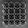
Calcium phosphate 3D lattice structures and patterns printed using the PNC ink. a) Models and SEM micrographs displaying a progression of 2PP-printed lattice structures from macro to nano scales. Structures include: Gyroid (optical image, 250 μm× 250 μm× 250 μm), Kelvin (100 μm× 100 μm× 25 μm), Cubic (30 μm × 30 μm × 12 μm), Voronoi (20 μm × 20 μm × 20 μm), FCC (5 μm × 5 μm × 5 μm) and Octet (unit cell size of 2 μm × 2 μm × 2 μm). b) Nanopatterning on opaque substrates: i) Microscale inscription of “Materials” and “Nano” next to a human hair (arrow) for scale. Each letter consists of arbitrary placed bicontinuous grains with consistent number of granular pockets in all patterns; ii) A large-area (500 μm × 500 μm) representation of Einstein’s face demonstrating surface roughness variations from 2.8 to 300 nm; iii) Rastered microporosity within a single “O” letter achieved by varying laser raster spacing; iv) An array of nano-needles showcasing a tip radius of 310 nm and base diameter of 25 μm; v) Uniform, single-cell tessellation with 6 μm diameters and double grain height; vi) Biomimetic sub-micron patterning emulating bone trabeculae. (Image: Reprinted from DOI:10.1002/adma.202413626, CC BY) (click on image to enlarge)
This clarity enables the use of two-photon polymerization, where precisely focused laser pulses solidify specific points within the printing material. The team achieved feature sizes of 300 nanometers – about 1/300th the width of a human hair. They demonstrated this capability by printing intricate structures including microscopic lattices with struts just 360 nanometers thick and arrays of precisely spaced mineral grains.
The technique’s precision allows recreation of bone’s internal features. The team printed artificial versions of osteons – the fundamental building units of bone – complete with concentric mineral rings and microscopic channels that mimic spaces for blood vessels. Additionally, they replicated trabecular bone microstructures and impact-resistant patterns found in lobster shells, demonstrating the method’s ability to mimic a variety of biological architectures. They also created nanoneedles, which could have applications in cellular manipulation and targeted drug delivery.
This level of structural control addresses fundamental limitations in artificial bone development. Current materials often fail because they lack the microscopic features that guide bone cell behavior and promote healing. The new printing method enables systematic investigation of how specific structural patterns influence cell response and bone regeneration. Moreover, precise nanoscale fabrication could help overcome the brittleness of traditional calcium phosphate materials, potentially enabling their use in load-bearing bone implants.
The technique also offers a path toward stronger artificial bone materials. Natural bone derives much of its strength from precise mineral organization at the nanoscale. By enabling similar control in synthetic structures, the method could help create materials that better match bone’s mechanical properties.
Two-photon polymerization-enabled CaP biomimetic structures. Biomimetic CaP structures fabricated using PNC ink. a) Engineered Bouligand structure designed to allow lamellar reorientation akin to load-responsivemechanisms observed in various naturalmaterials (scale bar, 10 μm). b) Scaled 1:20 of a micro bone tissue displaying osteons, Haversian channels, and concentric rings of oriented fibrils as well as an interface transition of nanothin trabecular bone to cortical bone, (scale bar, 10 μm). c) Scaled 1:100 representation of a histological section of a femur bone, demonstrating the larynx of trabecular bone with varying plate thickness (scale bar, 50 μm). (Image: Reprinted from DOI:10.1002/adma.202413626, CC BY)
Applications extend beyond bone replacement. The ability to print detailed mineral patterns on various surfaces could improve medical implant integration or enable new drug delivery systems. The researchers demonstrated this versatility by creating patterns on multiple materials including silicon, titanium alloys, and glass.
Several technical hurdles remain before clinical implementation. The printing process works slowly for larger structures, and the required high-temperature processing prevents direct incorporation of biological molecules during fabrication. However, the researchers demonstrated that biological components can be added afterward through surface modification techniques. Additionally, they suggest that hybrid structures combining calcium phosphate with biocompatible resins or hydrogels could allow for direct incorporation of biological molecules while maintaining structural integrity.
This advance in nanoscale mineral printing provides new tools for investigating and replicating the structural complexity of natural bone. The ability to precisely position calcium phosphate opens possibilities not only for improved bone replacement materials but also for broader applications in regenerative medicine and biomedical engineering.
Get our Nanotechnology Spotlight updates to your inbox!
Thank you!
You have successfully joined our subscriber list.
Become a Spotlight guest author! Join our large and growing group of guest contributors. Have you just published a scientific paper or have other exciting developments to share with the nanotechnology community? Here is how to publish on nanowerk.com.