Home > Press > Nanophotonics enhanced coverslip for phase imaging in biology
 |
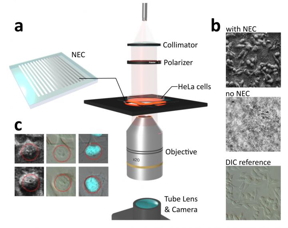
| a The nanophotonics enhanced coverslip (NEC) adds phase imaging capability to a normal microscope coverslip, thereby shrinking bulky phase-imaging methods down to the size of a chip. The less than 200 nm thick design consists of a subwavelength spaced grating on top of an optically thin film, supported by a glass substrate. b Exemplary demonstration of phase-imaging of human cancer cells (HeLa cells) using the NEC. By placing the Petri dish containing the cell culture directly on top of the NEC, pseudo 3D images of the cells are created. The obtained images are similar to those obtained by the conventional phase-imaging technique of differential interference contrast (DIC) microscopy. In the reference image, recorded without the NEC, the cells are mostly invisible. c Use of the NEC device not only enabled visualization of the general shape of the cell, but also features inside of the cell nucleus (left). This was confirmed via comparison with images obtained via conventional DIC microscopy (middle) and fluorescence microscopy (right).
CREDIT by Lukas Wesemann, Jon Rickett, Jingchao Song, Jieqiong Lou, Elizabeth Hinde, Timothy J. Davis, and Ann Roberts |
Abstract:
The ability to visualize transparent objects such as biological cells is of fundamental importance in biology and medical diagnostics. Conventional approaches to achieve this include phase-contrast microscopy and techniques that rely on chemical staining of biological cells. These techniques, however, rely on expensive and bulky optical components or require changing, and in some cases damaging, the cell by introducing chemical contrast agents. Significant recent advances in nanofabrication technology permit structuring materials on the nanoscale with unprecedented precision. This has given rise to the revolutionary field of meta-optics that aims to develop ultra-compact optical components that replace their bulk-optical counterparts as for example lenses and optical filters. Such meta-optical devices exhibit unusual properties for which they have recently drawn significant scientific interest as novel platforms for imaging applications.
Nanophotonics enhanced coverslip for phase imaging in biology
Changchun, China | Posted on May 14th, 2021
In a new paper published in Light Science & Applications, a team of scientists, led by Professor Ann Roberts from the University of Melbourne node of the Australian Research Council Center of Excellence for Transformative Meta-Optical Systems have developed an ultra-compact, nanostructured microscope coverslip that allows the visualization of unstained biological cells. The device is referred to as a nanophotonics enhanced coverslip (NEC) since it adds phase-imaging capability to a normal microscope coverslip. In their study the researchers demonstrated that by simply placing biological cells on top of the NEC, high-contrast pseudo 3D images of otherwise invisible cells are obtained. The scientists used the example of human cancer cells (HeLa cells) to demonstrate the potential of this new phase-imaging method. The method not only enabled visualization of the general shape of the cancer cells but also made details of the cell nucleus visible. This capacity is crucial since the detection of changes in the structure of biological cells underpins the detection of diseases as for example in the case of malaria.
The version of the NEC presented in the publication differs from a normal coverslip through the addition of a thin-optical film and a nanometer spaced grating. The research team, however, envisage more complex variations of this concept to further extend the capabilities of the method to operation at different wavelengths and integration into highly-specialized optical imaging or microfluidic systems. In conclusion, this research has demonstrated an entirely new phase-imaging method that carries significant potential to be part of future biological imaging systems and mobile medical diagnostic tools.
The scientists summarize the potential of their phase-imaging method:
“We designed a nanostructured microscope coverslip that allows us to visualize otherwise transparent biological cells simply by placing them on top of the device and shining light through them. This is an exciting breakthrough in the field of phase-imaging, since our method requires neither the use of bulk-optical components, chemical staining or computational post processing as it is the case with conventional methods.” Prof. Roberts explained.
“The unavailability of medical diagnostic tools in many developing nations is regarded a reason for infectious diseases like malaria and tuberculosis to still be a leading cause of death. Our approach has significant potential to become an inexpensive, ultra-compact phase-imaging tool that could be integrated into smartphone cameras and other mobile devices to make mobile medical diagnostics broadly available.” Dr. Wesemann added.
####
For more information, please click here
Contacts:
Ann Roberts
Copyright © Changchun Institute of Optics, Fine Mechanics and Physics, Chinese Academy of Sciences
If you have a comment, please Contact us.
Issuers of news releases, not 7th Wave, Inc. or Nanotechnology Now, are solely responsible for the accuracy of the content.
News and information
![]() Sensors developed at URI can identify threats at the molecular level: More sensitive than a dog’s nose and the sensors don’t get tired May 21st, 2021
Sensors developed at URI can identify threats at the molecular level: More sensitive than a dog’s nose and the sensors don’t get tired May 21st, 2021
![]() Luminaries: Steven DenBaars and John Bowers receive top recognition at Compound Semiconductor Week conference May 21st, 2021
Luminaries: Steven DenBaars and John Bowers receive top recognition at Compound Semiconductor Week conference May 21st, 2021
![]() New technology enables rapid sequencing of entire genomes of plant pathogens May 14th, 2021
New technology enables rapid sequencing of entire genomes of plant pathogens May 14th, 2021
Imaging
![]() Harvesting light like nature does:Synthesizing a new class of bio-inspired, light-capturing nanomaterials May 14th, 2021
Harvesting light like nature does:Synthesizing a new class of bio-inspired, light-capturing nanomaterials May 14th, 2021
![]() World’s first fiber-optic ultrasonic imaging probe for future nanoscale disease diagnostics April 30th, 2021
World’s first fiber-optic ultrasonic imaging probe for future nanoscale disease diagnostics April 30th, 2021
Govt.-Legislation/Regulation/Funding/Policy
![]() Sensors developed at URI can identify threats at the molecular level: More sensitive than a dog’s nose and the sensors don’t get tired May 21st, 2021
Sensors developed at URI can identify threats at the molecular level: More sensitive than a dog’s nose and the sensors don’t get tired May 21st, 2021
![]() New technology enables rapid sequencing of entire genomes of plant pathogens May 14th, 2021
New technology enables rapid sequencing of entire genomes of plant pathogens May 14th, 2021
![]() Harvesting light like nature does:Synthesizing a new class of bio-inspired, light-capturing nanomaterials May 14th, 2021
Harvesting light like nature does:Synthesizing a new class of bio-inspired, light-capturing nanomaterials May 14th, 2021
Possible Futures
![]() Sensors developed at URI can identify threats at the molecular level: More sensitive than a dog’s nose and the sensors don’t get tired May 21st, 2021
Sensors developed at URI can identify threats at the molecular level: More sensitive than a dog’s nose and the sensors don’t get tired May 21st, 2021
![]() Luminaries: Steven DenBaars and John Bowers receive top recognition at Compound Semiconductor Week conference May 21st, 2021
Luminaries: Steven DenBaars and John Bowers receive top recognition at Compound Semiconductor Week conference May 21st, 2021
![]() Harvesting light like nature does:Synthesizing a new class of bio-inspired, light-capturing nanomaterials May 14th, 2021
Harvesting light like nature does:Synthesizing a new class of bio-inspired, light-capturing nanomaterials May 14th, 2021
Discoveries
![]() Sensors developed at URI can identify threats at the molecular level: More sensitive than a dog’s nose and the sensors don’t get tired May 21st, 2021
Sensors developed at URI can identify threats at the molecular level: More sensitive than a dog’s nose and the sensors don’t get tired May 21st, 2021
![]() Luminaries: Steven DenBaars and John Bowers receive top recognition at Compound Semiconductor Week conference May 21st, 2021
Luminaries: Steven DenBaars and John Bowers receive top recognition at Compound Semiconductor Week conference May 21st, 2021
![]() Harvesting light like nature does:Synthesizing a new class of bio-inspired, light-capturing nanomaterials May 14th, 2021
Harvesting light like nature does:Synthesizing a new class of bio-inspired, light-capturing nanomaterials May 14th, 2021
Announcements
![]() Sensors developed at URI can identify threats at the molecular level: More sensitive than a dog’s nose and the sensors don’t get tired May 21st, 2021
Sensors developed at URI can identify threats at the molecular level: More sensitive than a dog’s nose and the sensors don’t get tired May 21st, 2021
![]() Luminaries: Steven DenBaars and John Bowers receive top recognition at Compound Semiconductor Week conference May 21st, 2021
Luminaries: Steven DenBaars and John Bowers receive top recognition at Compound Semiconductor Week conference May 21st, 2021
![]() Harvesting light like nature does:Synthesizing a new class of bio-inspired, light-capturing nanomaterials May 14th, 2021
Harvesting light like nature does:Synthesizing a new class of bio-inspired, light-capturing nanomaterials May 14th, 2021
Interviews/Book Reviews/Essays/Reports/Podcasts/Journals/White papers/Posters
![]() Sensors developed at URI can identify threats at the molecular level: More sensitive than a dog’s nose and the sensors don’t get tired May 21st, 2021
Sensors developed at URI can identify threats at the molecular level: More sensitive than a dog’s nose and the sensors don’t get tired May 21st, 2021
![]() New technology enables rapid sequencing of entire genomes of plant pathogens May 14th, 2021
New technology enables rapid sequencing of entire genomes of plant pathogens May 14th, 2021
![]() Harvesting light like nature does:Synthesizing a new class of bio-inspired, light-capturing nanomaterials May 14th, 2021
Harvesting light like nature does:Synthesizing a new class of bio-inspired, light-capturing nanomaterials May 14th, 2021
Tools
![]() World’s first fiber-optic ultrasonic imaging probe for future nanoscale disease diagnostics April 30th, 2021
World’s first fiber-optic ultrasonic imaging probe for future nanoscale disease diagnostics April 30th, 2021
![]() New Cypher VRS1250 Video-Rate Atomic Force Microscope Enables True Video-Rate Imaging at up to 45 Frames per Second April 30th, 2021
New Cypher VRS1250 Video-Rate Atomic Force Microscope Enables True Video-Rate Imaging at up to 45 Frames per Second April 30th, 2021
![]() Researchers realize high-efficiency frequency conversion on integrated photonic chip April 23rd, 2021
Researchers realize high-efficiency frequency conversion on integrated photonic chip April 23rd, 2021
![]() An easy-to-use platform is a gateway to AI in microscopy April 23rd, 2021
An easy-to-use platform is a gateway to AI in microscopy April 23rd, 2021
Grants/Sponsored Research/Awards/Scholarships/Gifts/Contests/Honors/Records
![]() Luminaries: Steven DenBaars and John Bowers receive top recognition at Compound Semiconductor Week conference May 21st, 2021
Luminaries: Steven DenBaars and John Bowers receive top recognition at Compound Semiconductor Week conference May 21st, 2021
![]() New brain-like computing device simulates human learning: Researchers conditioned device to learn by association, like Pavlov’s dog April 30th, 2021
New brain-like computing device simulates human learning: Researchers conditioned device to learn by association, like Pavlov’s dog April 30th, 2021
![]() Silver ions hurry up, then wait as they disperse: Rice chemists show ions staged release from gold-silver nanoparticles could be useful property April 23rd, 2021
Silver ions hurry up, then wait as they disperse: Rice chemists show ions staged release from gold-silver nanoparticles could be useful property April 23rd, 2021
Photonics/Optics/Lasers
![]() Luminaries: Steven DenBaars and John Bowers receive top recognition at Compound Semiconductor Week conference May 21st, 2021
Luminaries: Steven DenBaars and John Bowers receive top recognition at Compound Semiconductor Week conference May 21st, 2021
![]() Emergence of a new heteronanostructure library May 14th, 2021
Emergence of a new heteronanostructure library May 14th, 2021
![]() With new optical device, engineers can fine tune the color of light April 23rd, 2021
With new optical device, engineers can fine tune the color of light April 23rd, 2021
![]() Silver ions hurry up, then wait as they disperse: Rice chemists show ions staged release from gold-silver nanoparticles could be useful property April 23rd, 2021
Silver ions hurry up, then wait as they disperse: Rice chemists show ions staged release from gold-silver nanoparticles could be useful property April 23rd, 2021
Research partnerships
![]() Graphene key for novel hardware security May 10th, 2021
Graphene key for novel hardware security May 10th, 2021
![]() Silver ions hurry up, then wait as they disperse: Rice chemists show ions staged release from gold-silver nanoparticles could be useful property April 23rd, 2021
Silver ions hurry up, then wait as they disperse: Rice chemists show ions staged release from gold-silver nanoparticles could be useful property April 23rd, 2021
![]() TPU scientists offer new plasmon energy-based method to remove CO2 from atmosphere March 19th, 2021
TPU scientists offer new plasmon energy-based method to remove CO2 from atmosphere March 19th, 2021


