| Dec 19, 2023 |
|
(Nanowerk News) A group of researchers has created a simple and inexpensive means to visualize the atomic state of hydrogen.
|
|
Details of their breakthrough were published in the journal Acta Materialia (“In situ visualization of misorientation-dependent hydrogen diffusion at grain boundaries of pure polycrystalline Ni using a hydrogen video imaging system”).
|
|
Hydrogen is carbon dioxide free, and it has long been touted as a source of clean energy. Yet, shifting society towards a hydrogen energy-based one requires overcoming some significant technical issues. Structural and functional materials that produce, store, transport and preserve hydrogen are needed.
|
 |
| (a) Comparison of the spatial and time resolutions between the conventional hydrogen detection techniques and the hydrogen visualization technique developed in this study. (b) A schematic of the present hydrogen visualization technique. (Image: Hiroshi Kakinuma et al.)
|
|
To develop advanced materials for hydrogen-related applications, a fundamental understanding of how hydrogen behaves in alloys is crucial. However, current technology falls short in this area. Detecting atomic state hydrogen – the smallest atom in the universe – with X-rays or lasers is challenging due to its unique characteristics. Researchers are currently focusing on better analytical and visualization techniques that can incorporate high spatial and time resolutions simultaneously.
|
|
Hiroshi Kakinuma, an assistant professor at Tohoku University, and his co-authors developed a new visualization technique harnessing an optical microscope and polyaniline layer. “When the color of the polyaniline layer reacts with the atomic state hydrogen in metals, it changes colors, allowing us to analyze the flow of hydrogen atoms based on the color distribution of the polyaniline layer,” points out Kakinuma. “Additionally, optical microscopes can observe the sub-millimeter-scale view with microscale spatial resolution in real time, thereby capturing hydrogen behavior with unprecedented high spatial and time resolutions.”
|
|
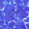
Thanks to this method, the researchers successfully filmed the flow of hydrogen atoms in pure nickel (Ni). The color of polyaniline changed from purple to white when reacting with hydrogen atoms in a metal. In situ visualization revealed that hydrogen atoms in pure Ni preferentially diffused through grain boundaries in disordered Ni atoms.
|
 |
| Optical micrographs of the polyaniline layer on pure Ni. Because hydrogen atoms diffuse in pure Ni with time, the flow of hydrogen atoms is visualized as a color change of the polyaniline layer (from purple to white). The color-changed area of the polyaniline layer corresponded to the grain boundaries of pure Ni, meaning that the grain boundaries are the preferential hydrogen diffusion path in pure Ni. (Image: Hiroshi Kakinuma et al.)
|
|
Furthermore, the group found that hydrogen diffusion was dependent on the geometrical structure of the grain boundaries: the hydrogen flux grew at grain boundaries with large geometric spaces. These results experimentally clarified the relationship between the atomic-scale structure of pure Ni and the hydrogen diffusion behavior.
|
|
The approach has broader applications as well. It can be applied to other metals and alloys, such as steels and aluminum alloys, and drastically facilitates elucidating the microscopic hydrogen-material interactions, which could be further investigated through simulations.
|
|
“Understanding hydrogen behaviors related to the atomic-scale structure of alloys will enable efficient alloy design, which will dramatically accelerate the development of highly functional materials and usher us one step closer to a hydrogen energy-based society,” adds Kakinuma.
|


