| Apr 22, 2025 |
Researchers successfully visualized how tiny structures inside one of these crystals respond to electric fields in real time – shedding light on the dynamics of nanostructure in materials used in ultrasound probes.
|
|
(Nanowerk News) Ultrasound imaging is one of the most widely used diagnostic tools in modern medicine. Behind its non-invasive magic lies a class of materials known as piezoelectric single crystals, which can convert electrical signals into mechanical vibrations and vice versa.
|
|
Now, in a world-first, a research team from Kumamoto University has successfully visualized how tiny structures inside one of these crystals respond to electric fields in real time—shedding light on the dynamics of nanostructure in materials used in ultrasound probes.
|
 |
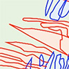
| Figure 1. Change of domain structure by AC poling. A transmission electron microscopy (TEM) image (top left) and the corresponding domain structure (bottom left) before applying an AC electric field. Top right and right bottom are TEM image and the corresponding domain structure after applying an AC electric field for 0.05 seconds. In the bottom panels, domain boundary is indicated by black and colored lines. Red and blue hatches in the right bottom panel indicate newly generated and laterally shrunk domains, respectively. Direction of the electric fields is shown in the top right panel. (Image: Courtesy of the researchers)
|
|
They published their findings in Applied Physics Letters (“Response of ferroelectric nanodomain to alternative-current electric fields in morphotropic-phase boundary Pb(Mg1/3Nb2/3)O3−PbTiO3“).
|
|
The team, led by Professor Yukio Sato from the Research and Education Institute for Semiconductors and Informatics (REISI), focused on a crystal known as PMN-PT (a solid solution of lead magnesium niobate and lead titanate), prized for its exceptional piezoelectric performance. It has been known that applying alternating current (AC) electric fields—known as AC poling—can enhance the performance of these materials. But the exact mechanisms behind this improvement, and how overuse can actually degrade performance, remained a mystery.
|
 |
| Figure 2. Change of domain structure by short-time AC poling. TEM images (top row) and the corresponding domain structures (bottom row) after applying AC electric fields for 0.05 seconds (left column) and 2 seconds (right column), respectively. (Image: Courtesy of the researchers)
|
|
To investigate, the team used a specialized in situ electron microscopy method developed at Kumamoto University, which allowed them to observe microscopic domain structures—called ferroelectric nanodomains—as they responded to AC electric fields.
|
|
What they saw was striking: just one cycle of an AC electric field at a strength of 12 kV/cm and 20 Hz significantly changed the domain structure (Figure 1). Over time, shorter AC treatments caused some domain walls to grow and merge, potentially enhancing the material’s properties (Figure 2). However, extended treatments led to the formation of vertically aligned microdomain bands that may hinder performance—a phenomenon consistent with over-poling (Figure 3).
|
 |
| Figure 3. Change of domain structure by long-time AC poling. TEM images (top row) and the corresponding domain structures (bottom row) after applying AC electric fields for 2 seconds (left column) and 870 seconds (right column), respectively. (Image: Courtesy of the researchers)
|
|
“This is the first time we’ve been able to watch these nanoscale domains react in real time,” says Professor Sato. “Understanding these changes is essential for refining the poling process and developing more efficient and longer-lasting medical imaging devices.”
|