
Home > Press > New microscope offers faster, high-resolution brain imaging: Enhanced two-photon microscopy method could reveal insights into neural dynamics and neurological diseases
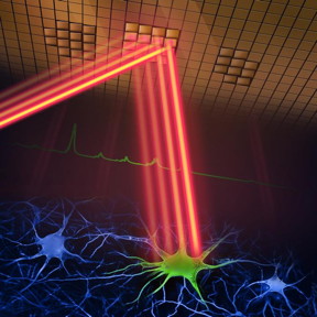 |
| A new two-photon fluorescence microscope can capture high-speed images of neural activity at cellular resolution thanks to a new adaptive sampling scheme and line illumination. The illustration shows the adaptive sampling scheme, in which a laser beam patterned by a digital micromirror device selectively illuminates neurons in the brain tissue to image their activity.
Credit Wei Wei and Mei Xueting, LINGO.AI LLC |
Abstract:
Researchers have developed a new two-photon fluorescence microscope that captures high-speed images of neural activity at cellular resolution. By imaging much faster and with less harm to brain tissue than traditional two-photon microscopy, the new approach could provide a clearer view of how neurons communicate in real time, leading to new insights into brain function and neurological diseases.
New microscope offers faster, high-resolution brain imaging: Enhanced two-photon microscopy method could reveal insights into neural dynamics and neurological diseases
Washington, DC | Posted on August 16th, 2024
Our new microscope is ideally suited for studying the dynamics of neural networks in real time, which is crucial for understanding fundamental brain functions such as learning, memory and decision-making, said research team leader Weijian Yang from the University of California, Davis. For example, researchers could use it to observe neural activity during learning to better understand communication and interaction among different neurons during this process.
In Optica, Optica Publishing Groups journal for high-impact research, the researchers describe the new two-photon fluorescence microscope, which incorporates a new adaptive sampling scheme and replaces traditional point illumination with line illumination. They show that the new method enables in vivo imaging of neuronal activity in a mouse cortex and can image at speeds ten times faster than traditional two-photon microscopy while also reducing the laser power on the brain more than tenfold.
By providing a tool that can observe neuronal activity in real time, our technology could be used to study the pathology of diseases at the earliest stages, said Yunyang Li, the first author of the paper. This could help researchers better understand and more effectively treat neurological diseases such as Alzheimers, Parkinsons and epilepsy.
High-speed imaging with less damage
Two-photon microscopy can image deep into scattering tissue such as a mouse brain by scanning a small point of light across the entire sample area to excite fluorescence and then collecting the resulting signal point by point. This process is then repeated to capture each imaging frame. Although two-photon microscopy provides detailed images, it is slow and can damage brain tissue.
In the new work, the researchers aimed to overcome these limitations through a new sampling strategy. Rather than using a point of light, they use a short line of light to illuminate specific parts of the brain where neurons are active. This enables a larger area to be excited and imaged at once, thus speeding up the imaging process significantly. Also, because it only images neurons of interest not the background or inactive areas the total light energy deposited to the brain tissue is reduced, lowering the risk for potential damage. They called this scheme adaptive sampling.
The researchers accomplished this by using a digital micromirror device (DMD) a chip containing thousands of tiny mirrors that can be individually controlled to dynamically shape and steer the light beam, enabling precise targeting of active neurons. They achieved adaptive sampling by turning individual DMD pixels on and off in a way that adjusts to the neuronal structure of the brain tissue being imaged.
The researchers also developed a technique to use the DMD to mimic high-resolution point scanning. This allows a high-resolution image to be reconstructed from fast scans, providing a quick way to identify neuronal regions of interest. This is critical for the subsequent high-speed imaging with the short-line excitation and adaptive sampling scheme.
These developments each crucial on its own come together to create a powerful imaging tool that significantly advances the ability to study dynamic neural processes in real time, with reduced risk to living tissue, said Yang. Importantly, our technique can be combined with other techniques like beam multiplexing and remote focusing to further increase the imaging speed or to achieve volumetric 3D imaging.
Capturing neural activity
The researchers demonstrated the new microscope by using it to image calcium signals indicators of neural activity in living mouse brain tissue. The system captured these signals at a speed of 198 Hz, which is significantly faster than traditional two-photon microscopes and demonstrates the ability to monitor rapid neuronal events that would be missed by slower imaging methods.
They also showed that the adaptive line-excitation technique coupled with advanced computational algorithms makes it possible to isolate the activity of individual neurons. This is important for accurately interpreting complex neural interactions and understanding the functional architecture of the brain.
Next, the researchers are working to integrate voltage imaging capabilities into the microscope to capture a direct and extremely rapid readout of neural activity. They also plan to use the new method for real neuroscience applications, such as observing neural activity during learning and studying brain activity in disease states. Additionally, they aim to improve the microscope’s user-friendliness and reduce its size to enhance its utility in neuroscience research.
####
About Optica
Optica is an open-access journal dedicated to the rapid dissemination of high-impact peer-reviewed research across the entire spectrum of optics and photonics. Published monthly by Optica Publishing Group, the Journal provides a forum for pioneering research to be swiftly accessed by the international community, whether that research is theoretical or experimental, fundamental or applied. Optica maintains a distinguished editorial board of more than 60 associate editors from around the world and is overseen by Editor-in-Chief Prem Kumar, Northwestern University, USA. For more information, visit Optica.
About Optica Publishing Group
Optica Publishing Group is a division of Optica, Advancing Optics and Photonics Worldwide. It publishes the largest collection of peer-reviewed content in optics and photonics, including 18 prestigious journals, the societys flagship member magazine, and papers from more than 835 conferences, including 6,500+ associated videos. With over 400,000 journal articles, conference papers and videos to search, discover and access, Optica Publishing Group represents the full range of research in the field from around the globe.
For more information, please click here
Contacts:
Media Contacts
Leah Poffenberger
Optica
Office: 2024161994
Aaron Cohen
Optica
Expert Contact
Weijian Yang
University of California, Davis
Copyright © Optica
If you have a comment, please Contact us.
Issuers of news releases, not 7th Wave, Inc. or Nanotechnology Now, are solely responsible for the accuracy of the content.
Bookmark:


News and information
![]()
Unveiling the power of hot carriers in plasmonic nanostructures August 16th, 2024
![]()
Faster than one pixel at a time new imaging method for neutral atomic beam microscopes developed by Swansea researchers August 16th, 2024
![]()
Physicists unlock the secret of elusive quantum negative entanglement entropy using simple classical hardware August 16th, 2024
![]()
Quantum pumping in molecular junctions August 16th, 2024
Imaging
![]()
Faster than one pixel at a time new imaging method for neutral atomic beam microscopes developed by Swansea researchers August 16th, 2024
Possible Futures
![]()
Groundbreaking precision in single-molecule optoelectronics August 16th, 2024
![]()
Physicists unlock the secret of elusive quantum negative entanglement entropy using simple classical hardware August 16th, 2024
![]()
Quantum pumping in molecular junctions August 16th, 2024
Discoveries
![]()
Groundbreaking precision in single-molecule optoelectronics August 16th, 2024
![]()
Physicists unlock the secret of elusive quantum negative entanglement entropy using simple classical hardware August 16th, 2024
![]()
Quantum pumping in molecular junctions August 16th, 2024
Announcements
![]()
Unveiling the power of hot carriers in plasmonic nanostructures August 16th, 2024
![]()
Faster than one pixel at a time new imaging method for neutral atomic beam microscopes developed by Swansea researchers August 16th, 2024
![]()
Researchers observe locked electron pairs in a superconductor cuprate August 16th, 2024
Interviews/Book Reviews/Essays/Reports/Podcasts/Journals/White papers/Posters
![]()
Researchers observe locked electron pairs in a superconductor cuprate August 16th, 2024
![]()
Groundbreaking precision in single-molecule optoelectronics August 16th, 2024
Tools
![]()
Faster than one pixel at a time new imaging method for neutral atomic beam microscopes developed by Swansea researchers August 16th, 2024
![]()
Single atoms show their true color July 5th, 2024
![]()
Atomic force microscopy in 3D July 5th, 2024
Photonics/Optics/Lasers
![]()
Groundbreaking precision in single-molecule optoelectronics August 16th, 2024
![]()
Single atoms show their true color July 5th, 2024
![]()
New method cracked for high-capacity, secure quantum communication July 5th, 2024