| Feb 28, 2025 |
Scientists have perfected a new technique for creating experimental movies of proteins in motion.
(Nanowerk News) Scientists have made enormous advances in understanding the structures of proteins over the past several decades. Imaging technologies like cryo-electron microscopy and x-ray crystallography help researchers visualize the shapes of proteins in unprecedented detail; however, these tools primarily produce static snapshots of molecules. To truly understand protein function, researchers need to see them in action.
|
|
Researchers at the University of Chicago and Argonne National Laboratory have been working on this problem for years; now, together with partners from Harvard University, they have perfected a new technique for creating experimental movies of proteins in motion.
|
|
In a paper published recently in Cell (“Direct visualization of electric-field-stimulated ion conduction in a potassium channel”), they demonstrated the method, called electric-field stimulated time-resolved X-ray crystallography (EFX), on a potassium ion channel, a pore in the cell membrane that regulates the movement of potassium in and out of cells. The resulting videos confirmed findings from other research over the past 25 years using much more painstaking biochemical approaches, showing that EFX can be a powerful new tool for quickly visualizing and understanding protein dynamics.
|
|
“The fundamental problem is that we’ve never had methods for simple experiments to see proteins in motion, because proteins are really small and they move really fast,” said Rama Ranganathan, PhD, one of the senior authors of the new study. “But the future of structural biology is going to be in looking at the mechanics, or the dynamics of molecules. So, I think what we’ve done here is deliver a technology that can get us there.”
|
The magical experiment
|
|
Ranganathan, who is the Joseph Regenstein Professor in the Department of Biochemistry and Molecular Biology and the Pritzker School of Molecular Engineering at UChicago, began this work when he was at the University of Texas Southwestern Medical Center.
|
|
In 2016, his team published a paper in Nature (“Electric-field-stimulated protein mechanics”) first describing how they used an electrical field to make proteins move while they captured images using time resolved crystallography. This approach takes crystallized version of the protein of interest and places it in the path of an x-ray beam. When the x-rays strike the crystal, they are scattered in many directions, producing patterns that can be analyzed to produce the full three-dimensional shape. The “time resolved” part of this technique means that they take continuous images of the protein as it undergoes structural changes, capturing it in motion.
|
|
“It was the magical experiment that we’ve all been waiting for,” Ranganathan said, but the process was difficult and time consuming. He moved to UChicago in 2017, largely for the opportunity to direct the BioCARS facility, which provides access to state-of-the-art x-ray technologies using the Advanced Photon Source at Argonne National Laboratory. His team continued studying protein dynamics, among other things, working together with the new study’s other senior author Doeke Hekstra, PhD, a former postdoc in Ranganathan’s lab who is now an Associate Professor of Molecular and Cellular Biology and Applied Physics at Harvard.
|
|
As they perfected the EFX technology, they focused on studying the potassium ion channel, a fundamental cellular structure that is involved in a host of biological processes. Importantly, the activity of the ion channel is initiated by applying an electrical field to make ions move back and forth through the pore. This way, the team could experimentally manipulate the action to record the activity they wanted to see.
|
|
As they tracked ions flowing through the channel, they also saw unique mechanical features of the channel at work that matched with different observations collected over years by other researchers. The difference is that those scientists had to use time-consuming, labor-intensive methods like inducing genetic mutations to manipulate the channels, whereas EFX was able to capture ion channel activity in one neat video.
|
|
“Over the time scale of a couple nanoseconds, we were able to see ions flowing through the pore of this channel,” Ranganathan said. “All of those 25 years of knowledge, we could see it in the dynamics of one channel during its operation.”
|
 |
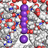
| An experimental video created using electric-field stimulated time-resolved X-ray crystallography (EFX) captures molecules moving through a potassium ion channel. (Animation: University of Chicago)
|
|
The goal is to integrate these dynamic observations with computational models to enhance protein engineering and design. Ranganathan is already deeply involved in using AI to design and build new custom proteins, through his biotech company Evozyne. Its platform simulates and learns from models of millions of years of evolution to create new proteins for specific purposes, like antibodies to treat disease or for capturing carbon to minimize industrial emissions. Detailed video images of proteins in action—real or simulated—could be incorporated into these models to refine and improve on these designs.
|
|
“We could create a virtuous cycle between computational prediction and experiment, so we can refine our simulations to the point where they truly look like the experiments,” Ranganathan said. “Once we unleash the simulations on all the molecules where people have solved the structure, we could build a database of dynamics with which we can really make computational predictions of all proteins in action.”
|