| Feb 02, 2024 |
|
|
|
(Nanowerk News) A team of University of Wisconsin–Madison scientists has developed the first 3D-printed brain tissue that can grow and function like typical brain tissue.
|
|
It’s an achievement with important implications for scientists studying the brain and working on treatments for a broad range of neurological and neurodevelopmental disorders, such as Alzheimer’s and Parkinson’s disease.
|
|
“This could be a hugely powerful model to help us understand how brain cells and parts of the brain communicate in humans,” says Su-Chun Zhang, professor of neuroscience and neurology at UW–Madison’s Waisman Center. “It could change the way we look at stem cell biology, neuroscience, and the pathogenesis of many neurological and psychiatric disorders.”
|
|
Printing methods have limited the success of previous attempts to print brain tissue, according to Zhang and Yuanwei Yan, a scientist in Zhang’s lab. The group behind the new 3D-printing process described their method in the journal Cell Stem Cell (“3D bioprinting of human neural tissues with functional connectivity”).
|
|
Instead of using the traditional 3D-printing approach, stacking layers vertically, the researchers went horizontally. They situated brain cells, neurons grown from induced pluripotent stem cells, in a softer “bio-ink” gel than previous attempts had employed.
|
|
“The tissue still has enough structure to hold together but it is soft enough to allow the neurons to grow into each other and start talking to each other,” Zhang says.
|
|
The cells are laid next to each other like pencils laid next to each other on a tabletop.
|
|
“Our tissue stays relatively thin and this makes it easy for the neurons to get enough oxygen and enough nutrients from the growth media,” Yan says.
|
|
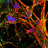
The results speak for themselves — which is to say, the cells can speak to each other. The printed cells reach through the medium to form connections inside each printed layer as well as across layers, forming networks comparable to human brains. The neurons communicate, send signals, interact with each other through neurotransmitters, and even form proper networks with support cells that were added to the printed tissue.
|
|
“We printed the cerebral cortex and the striatum and what we found was quite striking,” Zhang says. “Even when we printed different cells belonging to different parts of the brain, they were still able to talk to each other in a very special and specific way.”
|
|
The printing technique offers precision — control over the types and arrangement of cells — not found in brain organoids, miniature organs used to study brains. The organoids grow with less organization and control.
|
|
“Our lab is very special in that we are able to produce pretty much any type of neurons at any time. Then we can piece them together at almost any time and in whatever way we like,” Zhang says. “Because we can print the tissue by design, we can have a defined system to look at how our human brain network operates. We can look very specifically at how the nerve cells talk to each other under certain conditions because we can print exactly what we want.”
|
|
That specificity provides flexibility. The printed brain tissue could be used to study signaling between cells in Down syndrome, interactions between healthy tissue and neighboring tissue affected by Alzheimer’s, testing new drug candidates, or even watching the brain grow.
|
|
“In the past, we have often looked at one thing at a time, which means we often miss some critical components. Our brain operates in networks. We want to print brain tissue this way because cells do not operate by themselves. They talk to each other. This is how our brain works and it has to be studied all together like this to truly understand it,” Zhang says. “Our brain tissue could be used to study almost every major aspect of what many people at the Waisman Center are working on. It can be used to look at the molecular mechanisms underlying brain development, human development, developmental disabilities, neurodegenerative disorders, and more.”
|
|
The new printing technique should also be accessible to many labs. It does not require special bio-printing equipment or culturing methods to keep the tissue healthy, and can be studied in depth with microscopes, standard imaging techniques and electrodes already common in the field.
|
|
The researchers would like to explore the potential of specialization, though, further improving their bio-ink and refining their equipment to allow for specific orientations of cells within their printed tissue.
|
|
“Right now, our printer is a benchtop commercialized one,” Yan says. “We can make some specialized improvements to help us print specific types of brain tissue on-demand.”
|

