| Oct 13, 2023 |
|
|
|
(Nanowerk News) Acoustic radiation force generated by ultrasonic standing wave is one of the forces with the ability of cell trapping. Cells can be trapped at either pressure nodes or anti-nodes depending on the properties of the cells and suspension medium.
|
|
A research team from the Suzhou Institute of Biomedical Engineering and Technology (SIBET) of the Chinese Academy of Sciences has developed an acoustic trapping chip that can provide three-dimensional (3D) trapping of cells in a continuously flowing medium with a circular resonance structure.
|
|
The findings have been published in Sensors and Actuators A: Physical (“Acoustic 3D trapping of microparticles in flowing liquid using circular cavity”).
|
 |
| Schematic diagram of the chip design and its trapping performance on red blood cells and white blood cells. The cells aggregate to the center of the circular cavity within 60 ms. (Image: SIBET)
|
|
Cell trapping is of great importance in biomedical engineering because it enables the clamping, separation, filtration and agglomeration of cells. Among different trapping approaches, acoustic trapping has been widely used in biological research because it can provide contactless and biosafe cell manipulation.
|
|
Ultrasonic standing waves can be further categorized into standing bulk acoustic waves (BAW), generated by a bulk piezoelectric transducer, or standing surface acoustic waves (SAW), generated by single crystal lithium niobate (LiNbO3) etched with interdigitated electrodes. SAW can manipulate particles with very low energy consumption, but it is generally used for sorting in flowing liquid and particle arrangement in stationary liquid due to its overall smaller clamping force compared with BAW.
|
 |
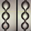
| Single particle with diameter of 10 μm queuing up on the cavity array was delivered step by step to the detection spot, which can be applied to high throughput Reman spectra acquisition. (Image: SIBET)
|
|
On the other hand, the acoustic microstreaming vortex can also be applied to trap cells near the obstacle or microbubbles. The design of micropillars or obstacles plays an important role in improving the trapping efficiency. However, some of the traps cannot release particles easily, and some of them cannot provide a fixed trapping position.
|
|
The trapping efficiency is basically determined by the trapping force. In most previous studies, particles are usually trapped in a static fluid or a fluid flowing at extremely low speed, or the trapping process takes several seconds, which is mainly due to insufficient trapping force. This reduces both trapping efficiency and throughput, while high-throughput cell manipulation is important in many biological applications, such as Raman identification and nanoparticle capture.
|
 |
| Chip used for nanoparticle capture. (a). Bright field image of seed cluster before nanoparticle capture; 10 μm blank polystyrene beads aggregate in the circular cavity; (b). Fluorescent image of seed cluster; As the seed particles are blank, nothing can be seen. (c). Fluorescent image of 100 nm green fluorescent nanoparticles captured in the region of the seed cluster. (Image: SIBET)
|
|
The researchers established a standing acoustic wave in the circular microstructure, providing sufficient force to clamp cells in the center of the chamber. Meanwhile, cells near the bottom of the microfluidic channel are clamped under the radiation force generated in the depth direction.
|
|
Thus, a 3D cell confinement is formed with a special design of microchannels actuated by only one piezoelectric plate transducer. Experimental results show that the chip can provide nanonewton (nN) level trapping force and millisecond (ms) level trapping time for micron-sized particles moving at the speed of mm/s level.
|
|
With this non-contact and biocompatible trapping method, the chip can be applied to a variety of biomedical engineering scenarios such as organ chips, cell culture, Raman analysis and nanoparticle capture.
|


