| Mar 25, 2022 |
|
(Nanowerk News) Most conventional microfluidic devices are fabricated in inherently planar, block-like devices that can’t be changed once fabricated. In contrast, an important feature of naturally self-assembled systems such as leaves and tissues is that they are curved and have embedded fluidic channels that enable the transport of nutrients to, or removal of waste from, specific three-dimensional regions.
|
|
To apply microfluidics in the biomedical field, such as the investigation of disease models, tissue development, and drug screening, it would ideally require three-dimensional (3D) microfluidic structures that can mimic the biological vascular networks in the human body.
|
|
Addressing this issue and opening new design possibilities for microfluidics, researchers have developed shape-programmable 3D microfluidic structures, which are assembled from a bilayer of channel-embedded polydimethylsiloxane (PDMS) and shape-memory polymers (SMPs) via compressive buckling.
|
|
They report their findings in ACS Applied Materials & Interfaces (“Shape-Programmable Three-Dimensional Microfluidic Structures”).
|
|
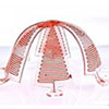
The researchers assembled a novel type of shape-programmable, open-mesh 3D microfluidic structures via compressive buckling of 2D PDMS/SMP precursors. They fabricated microfluidic channels in the PDMS layer via soft lithography and then laminated onto an SMP film to form a bilayer of PDMS/SMP, which is patterned into desired geometries and compressively buckled into 3D microfluidics by using a pre-stretched elastomer as an assembly platform.
|
 |
| Figure 1. Shape-programmable 3D microfluidic structures formed from a bilayer of PDMS/SMPs. (A) Schematic illustration of fabricating shape-programmable 3D microfluidic structures via a compressive buckling technique. (B) Optical images of various shape-programmable 3D microfluidic structures including their magnified views. Scale bars: 3 mm. (Reprinted with permission by American Chemical Society) (click on image to enlarge)
|
|
The incorporated shape-memory polymers allow the 3D microfluidic structures to be programmed into temporary, freestanding shapes and then recover their original shape under thermal stimuli and external forces.
|
|
The researchers point out that fluid circulation of the microfluidic channels is well maintained in both deformed and recovered shapes of the structures.
|
|
As illustrated in Figure 2, the team also demonstrated that by integrating magnetic particles into PDMS, fast, remote shape programming of 3D microfluidics via a portable magnet can be realized. The thicknesses of the SMP and PDMS layers for all structures in Figure 2 are 130 and 110 µm, respectively. SMPs can be programmed from an original (permanent) shape to a deformed (temporary) shape under external forces when exposed to external stimuli, such as heat or light.
|
 |
| Figure 2. Shape-memory cycle of shape-programmable 3D microfluidic structures. Diluted red food dye is injected into microfluidic channels for demonstration. (a) “Ribbon” structure. (b) “Stadium” structure. (c) “Table” structure. (d) “Umbrella” structure. (e) “Basket” structure. (f) “Box” structure, with optical and SEM images of the cross section of the SMP/PDMS interface. Scale bars for buckled structures a−f: 3 mm. Scale bars in the magnified views of structure f are 250, 100, and 250 µm from left to right. (Reprinted with permission by American Chemical Society)
|
|
The results provide significant guidance for the design and development of programmable 3D microfluidics for many applications including remotely controlled biomedical robotics, drug delivery systems, and artificial blood vessels.
|


